Skip to content

Lunaphore COMET

Sirona Dx is the first CRO in North America to provide access to the COMET platform from Lunaphore.

Our industry leading spatial biology service suite now includes the COMET platform to perform hyperplex assays based on flexible and fully automated sequential immunofluorescence (seqIF) to develop custom panels.

Capabilities

  • Throughput – high capacity (4 slides) and rapid turnaround time (40 minutes every two markers) provide unmatched hyperplex throughput.
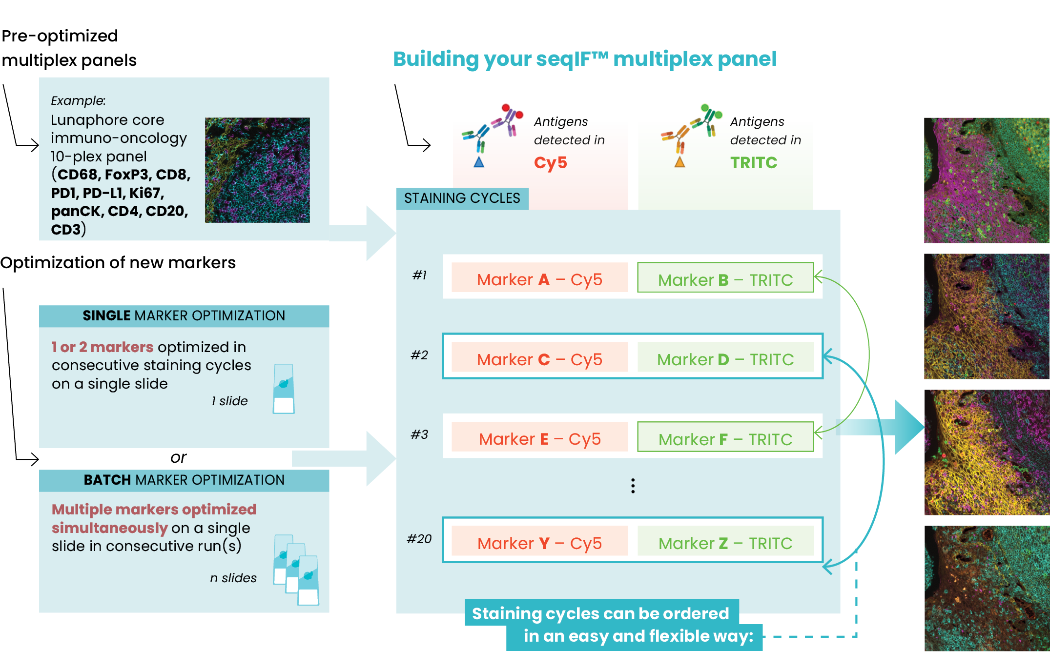
  • Fast assay development: panels can be developed in weeks rather than months. Panels are optimized by characterizing simple modules of 1-2 markers. Once characterized, markers can be shuffled in any protocol cycle or added to any new panel without any further optimization. Batch optimization possible (Figure 2).
  • Hyperplex: automated 40-plex runs detected on the same tissue slide.
  • Reproducibility – full automation, use of standard primary antibodies without the need of complex upstream conjugation and direct detection via secondary antibodies, eliminate downstream complexity and variability leading to high reproducibility.
  • Quality – highly controlled staining conditions thanks to a microfluidic technology (FFeX™) deliver immunofluorescence multiplex analysis comparable to classic IHC.
  • Preserved tissue morphology – low exposure to reagents thanks to rapid incubations, and a gentle antibody removal, preserve the tissue to obtain high-quality stainings and enable downstream applications and H&E cross-validation on the same sample.
  • Off-the-shelf primary antibodies – with a reagent agnostic approach, optimize using your own clones and reagent library; no conjugation and further validation required.
  • Subcellular resolution – 230 nm resolution
  • Integrated microscope – 20X magnification, TRITC, Cy5, DAPI channels.
  • Common sample types: FFPE, FS, TMAs
  • Output OME.TIFF – file supported by most image analysis tools.

Content

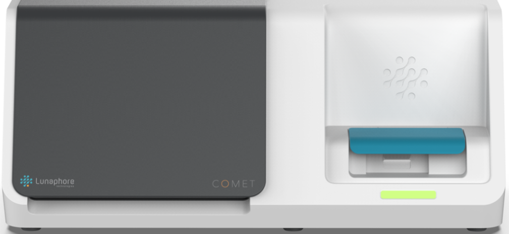
lunaphore-comet-front-new

Our industry leading spatial biology service suite now includes the COMET platform to perform hyperplex assays based on flexible and fully automated sequential immunofluorescence (seqIF) to develop custom panels.

Capabilities

  • Throughput – high capacity (4 slides) and rapid turnaround time (40 minutes every two markers) provide unmatched hyperplex throughput.
  • Fast assay development: panels can be developed in weeks rather than months. Panels are optimized by characterizing simple modules of 1-2 markers. Once characterized, markers can be shuffled in any protocol cycle or added to any new panel without any further optimization. Batch optimization possible (Figure 2).
  • Hyperplex: automated 40-plex runs detected on the same tissue slide.
  • Reproducibility – full automation, use of standard primary antibodies without the need of complex upstream conjugation and direct detection via secondary antibodies, eliminate downstream complexity and variability leading to high reproducibility.
  • Quality – highly controlled staining conditions thanks to a microfluidic technology (FFeX™) deliver immunofluorescence multiplex analysis comparable to classic IHC.
  • Preserved tissue morphology – low exposure to reagents thanks to rapid incubations, and a gentle antibody removal, preserve the tissue to obtain high-quality stainings and enable downstream applications and H&E cross-validation on the same sample.
  • Off-the-shelf primary antibodies – with a reagent agnostic approach, optimize using your own clones and reagent library; no conjugation and further validation required.
  • Subcellular resolution – 230 nm resolution
  • Integrated microscope – 20X magnification, TRITC, Cy5, DAPI channels.
  • Common sample types: FFPE, FS, TMAs
  • Output OME.TIFF – file supported by most image analysis tools.

Content

Building seqIF panels

Figure 1. Hyperplex panels can be easily developed on COMET with guided protocols for optimization and flexible choice of marker positioning.
COMET single-cell resolution

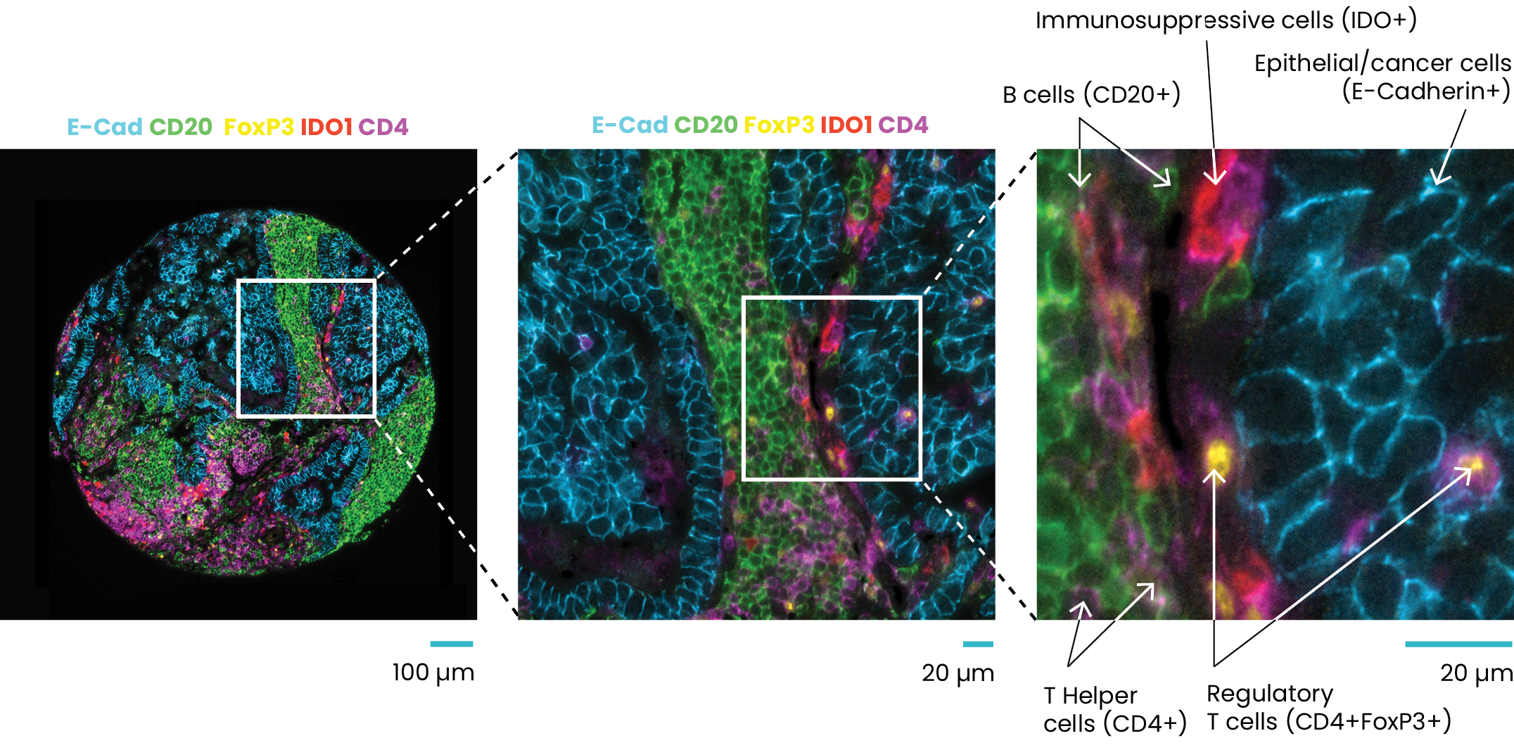
Figure 3. Single-cell resolution achieved with 40-plex staining on COMET. The left panel shows five markers detected on a TMA of lung adenocarcinoma with lymph node metastasis. The middle and right panels show a selection of regions of interest (ROIs) to identify different cell types.

Comprehensive End to End Service Includes:

  • Assay customization
  • Multiplex Assay Optimization
  • Slide Staining
  • Advanced Bioinformatics Analysis

Applications

  • Biomarker discovery and development
  • Immuno-oncology
  • Inflammatory and autoimmune diseases
  • Neuro-immunology
  • Characterization of tissue microenvironment
  • Therapeutic response
  • Drug safety and toxicity
  • Oncology

Please reach us at [email protected] for more information

Contact Us

Address

4640 Southwest Macadam Avenue,
Portland, Oregon 97239

Hours

Monday – Friday: 8:00am – 6:00pm PST
Saturday – Sunday: Closed